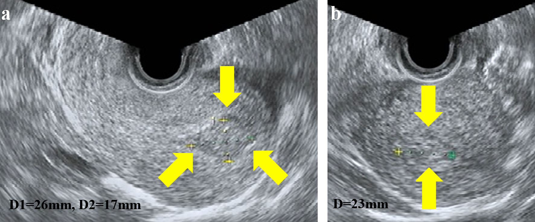
Figure 1. Transvaginal ultrasonography showed a 26 × 17 mm hyperechoic mass in the uterus; sagittal plane (a) and transverse plane (b).
| Journal of Clinical Gynecology and Obstetrics, ISSN 1927-1271 print, 1927-128X online, Open Access |
| Article copyright, the authors; Journal compilation copyright, J Clin Gynecol Obstet and Elmer Press Inc |
| Journal website https://www.jcgo.org |
Case Report
Volume 12, Number 3, December 2023, pages 117-122
Sal-Like Protein 4-Negative Gestational Trophoblastic Neoplasia
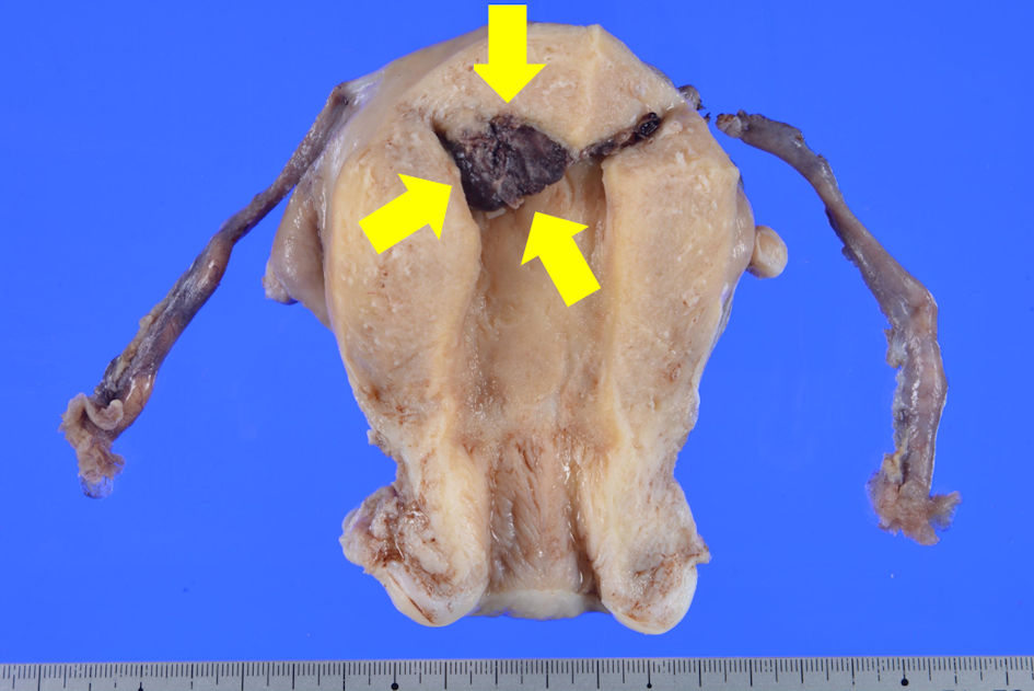
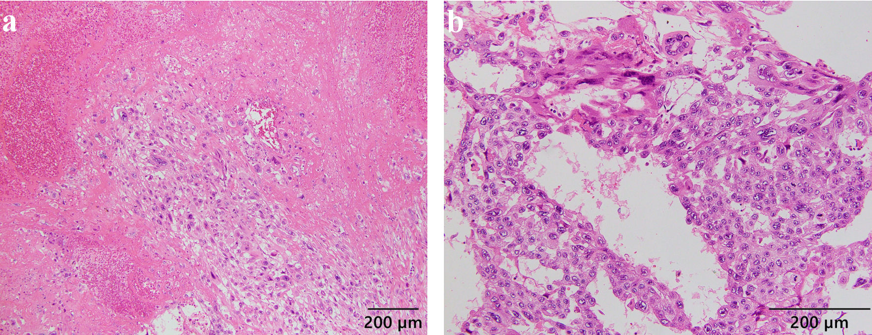
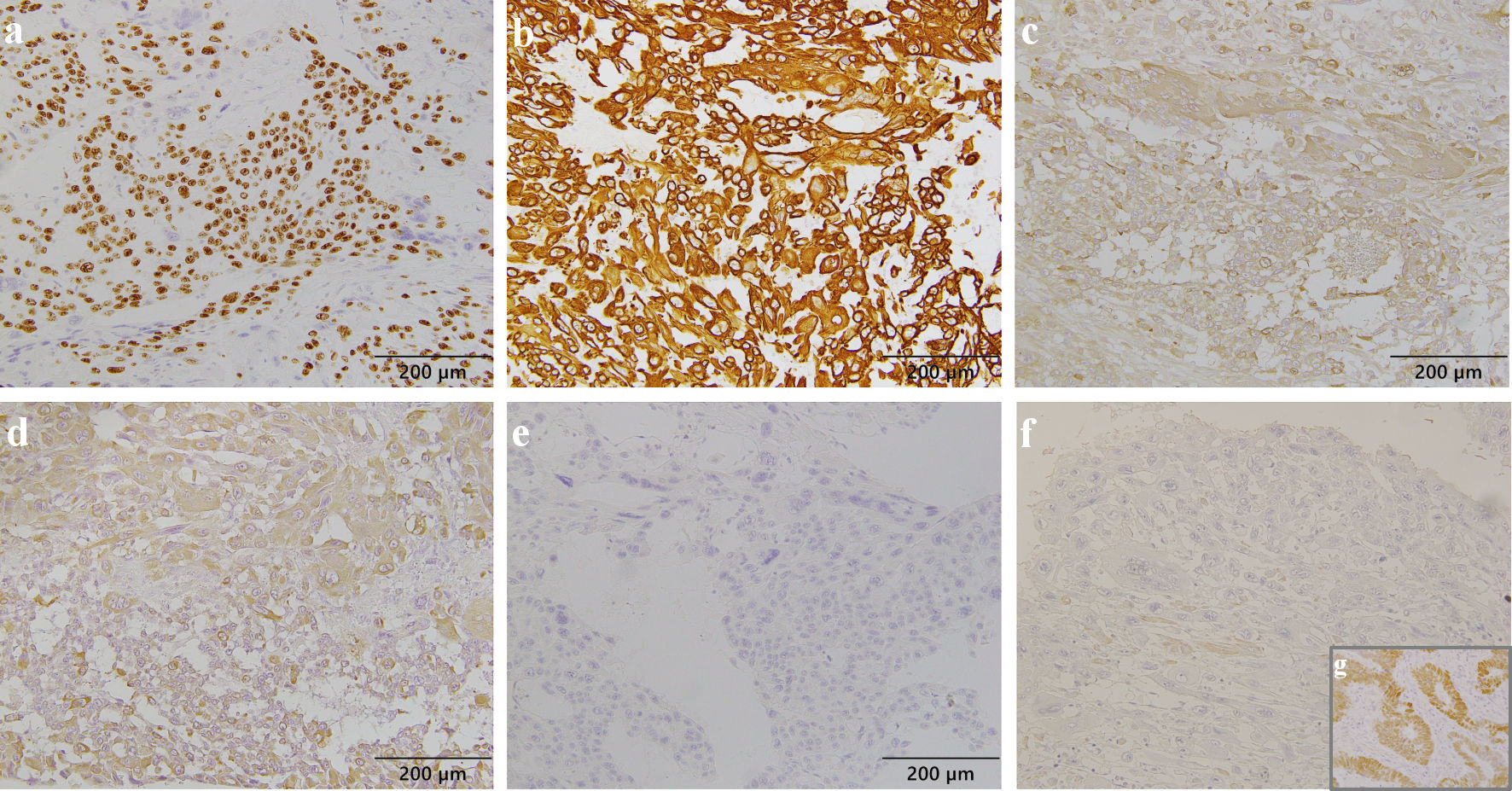
Figures